Figure 1
Painful unilateral gynecomastia with identification of the cause of the pain: A case report
Charles M Lombard* and Peter L Naruns
Published: 31 December, 2021 | Volume 5 - Issue 1 | Pages: 034-036

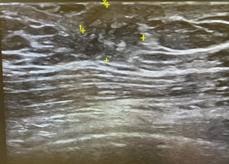
Figure 1:
Ultrasound of breast lesion. Ultrasound shows a hypoechoic rounded area in the subareolar tissues.
Read Full Article HTML DOI: 10.29328/journal.apcr.1001026 Cite this Article Read Full Article PDF
More Images
Similar Articles
-
MicroRNA Therapeutics in Triple Negative Breast CancerSarmistha Mitra*. MicroRNA Therapeutics in Triple Negative Breast Cancer . . 2017 doi: 10.29328/journal.hjpcr.1001003; 1: 009-017
-
A great mimicker of Bone Secondaries: Brown Tumors, presenting with a Degenerative Lumber Disc like painZuhal Bayramoglu*,Ravza Yılmaz,Aysel Bayram. A great mimicker of Bone Secondaries: Brown Tumors, presenting with a Degenerative Lumber Disc like pain. . 2017 doi: 10.29328/journal.hjpcr.1001004; 1: 018-023
-
Amyotropyc Lateral Sclerosis and Endogenous -Esogenous Toxicological Movens: New model to verify other Pharmacological StrategiesMauro Luisetto*,Behzad Nili-Ahmadabadi,Nilesh M Meghani,Ghulam Rasool Mashori,Ram Kumar Sahu,Kausar Rehman Khan, Ahmed Yesvi Rafa,Luca Cabianca,Gamal Abdul Hamid, Farhan Ahmad Khan. Amyotropyc Lateral Sclerosis and Endogenous -Esogenous Toxicological Movens: New model to verify other Pharmacological Strategies. . 2018 doi: 10.29328/journal.apcr.1001009; 2: 029-048
-
Receptor pharmacology and other relevant factors in lower urinary tract pathology under a functional and toxicological approach: Instrument to better manage antimicrobials therapyMauro Luisetto*,Naseer Almukhtar,Behzad Nili-Ahmadabadi,Ghulam Rasool Mashori,Kausar Rehman Khan,Ram Kumar Sahu,Farhan Ahmad Khan,Gamal Abdul Hamid,Luca Cabianca. Receptor pharmacology and other relevant factors in lower urinary tract pathology under a functional and toxicological approach: Instrument to better manage antimicrobials therapy . . 2018 doi: 10.29328/journal.apcr.1001010; 2: 049-093
-
The pathogenesis of psoriasis: insight into a complex “Mobius Loop” regulation processYuankuan Jiang,Haiyang Chen,Jiayue Liu,Tianfu Wei,Peng Ge,Jialin Qu*,Jingrong Lin. The pathogenesis of psoriasis: insight into a complex “Mobius Loop” regulation process. . 2021 doi: 10.29328/journal.apcr.1001024; 5: 020-025
-
Painful unilateral gynecomastia with identification of the cause of the pain: A case reportCharles M Lombard*,Peter L Naruns. Painful unilateral gynecomastia with identification of the cause of the pain: A case report. . 2021 doi: 10.29328/journal.apcr.1001026; 5: 034-036
-
Immune-mediated neuropathy related to bortezomib in a patient with multiple myelomaSusanne Koeppen*,Jörg Hense,Kay Wilhelm Nolte,Joachim Weis. Immune-mediated neuropathy related to bortezomib in a patient with multiple myeloma. . 2022 doi: 10.29328/journal.apcr.1001028; 6: 001-004
-
Post-operative agranulocytosis caused by intravenous cefazolin: A case report with a discussion of the pathogenesisCharles M Lombard*,Jiali Li,Bijayee Shrestha. Post-operative agranulocytosis caused by intravenous cefazolin: A case report with a discussion of the pathogenesis. . 2022 doi: 10.29328/journal.apcr.1001030; 6: 009-012
-
Harmonizing Artificial Intelligence Governance; A Model for Regulating a High-risk Categories and Applications in Clinical Pathology: The Evidence and some ConcernsMaxwell Omabe*. Harmonizing Artificial Intelligence Governance; A Model for Regulating a High-risk Categories and Applications in Clinical Pathology: The Evidence and some Concerns. . 2024 doi: 10.29328/journal.apcr.1001040; 8: 001-005
-
The Accuracy of pHH3 in Meningioma Grading: A Single Institution StudyMansouri Nada1, Yaiche Rahma*, Takout Khouloud, Gargouri Faten, Tlili Karima, Rachdi Mohamed Amine, Ammar Hichem, Yedeas Dahmani, Radhouane Khaled, Chkili Ridha, Msakni Issam, Laabidi Besma. The Accuracy of pHH3 in Meningioma Grading: A Single Institution Study. . 2024 doi: 10.29328/journal.apcr.1001041; 8: 006-011
Recently Viewed
-
Seismic Analysis of Semi-rigid Frames using Skeleton JointsCai Zi,Sun Menghan*,Zhang Chunhui,He luyao,Yang Zailin. Seismic Analysis of Semi-rigid Frames using Skeleton Joints. Ann Civil Environ Eng. 2025: doi: 10.29328/journal.acee.1001083; 9: 073-079
-
Scientific Analysis of Eucharistic Miracles: Importance of a Standardization in EvaluationKelly Kearse*,Frank Ligaj. Scientific Analysis of Eucharistic Miracles: Importance of a Standardization in Evaluation. J Forensic Sci Res. 2024: doi: 10.29328/journal.jfsr.1001068; 8: 078-088
-
Transnasal Humidified Rapid-Insufflation Ventilatory Exchange (THRIVE) in High-Risk Endoscopic Procedures: A Non-Intubation ApproachNaba Madoo*,Vaibhavi Baxi. Transnasal Humidified Rapid-Insufflation Ventilatory Exchange (THRIVE) in High-Risk Endoscopic Procedures: A Non-Intubation Approach. Int J Clin Anesth Res. 2025: doi: 10.29328/journal.ijcar.1001033; 9: 035-036
-
A Strength-based Approach to Achieving Academic Success for Individuals with Autism Spectrum Disorder (ASD)Sally M Reis*, Nicholas Gelbar, Joseph Madaus. A Strength-based Approach to Achieving Academic Success for Individuals with Autism Spectrum Disorder (ASD). J Neurosci Neurol Disord. 2024: doi: 10.29328/journal.jnnd.1001090; 8: 010-011
-
Why is Pain not Characteristic of Inflammation of the Lung Tissue?Igor Klepikov*. Why is Pain not Characteristic of Inflammation of the Lung Tissue?. J Clin Intensive Care Med. 2024: doi: 10.29328/journal.jcicm.1001046; 9: 005-007
Most Viewed
-
Feasibility study of magnetic sensing for detecting single-neuron action potentialsDenis Tonini,Kai Wu,Renata Saha,Jian-Ping Wang*. Feasibility study of magnetic sensing for detecting single-neuron action potentials. Ann Biomed Sci Eng. 2022 doi: 10.29328/journal.abse.1001018; 6: 019-029
-
Evaluation of In vitro and Ex vivo Models for Studying the Effectiveness of Vaginal Drug Systems in Controlling Microbe Infections: A Systematic ReviewMohammad Hossein Karami*, Majid Abdouss*, Mandana Karami. Evaluation of In vitro and Ex vivo Models for Studying the Effectiveness of Vaginal Drug Systems in Controlling Microbe Infections: A Systematic Review. Clin J Obstet Gynecol. 2023 doi: 10.29328/journal.cjog.1001151; 6: 201-215
-
Prospective Coronavirus Liver Effects: Available KnowledgeAvishek Mandal*. Prospective Coronavirus Liver Effects: Available Knowledge. Ann Clin Gastroenterol Hepatol. 2023 doi: 10.29328/journal.acgh.1001039; 7: 001-010
-
Causal Link between Human Blood Metabolites and Asthma: An Investigation Using Mendelian RandomizationYong-Qing Zhu, Xiao-Yan Meng, Jing-Hua Yang*. Causal Link between Human Blood Metabolites and Asthma: An Investigation Using Mendelian Randomization. Arch Asthma Allergy Immunol. 2023 doi: 10.29328/journal.aaai.1001032; 7: 012-022
-
An algorithm to safely manage oral food challenge in an office-based setting for children with multiple food allergiesNathalie Cottel,Aïcha Dieme,Véronique Orcel,Yannick Chantran,Mélisande Bourgoin-Heck,Jocelyne Just. An algorithm to safely manage oral food challenge in an office-based setting for children with multiple food allergies. Arch Asthma Allergy Immunol. 2021 doi: 10.29328/journal.aaai.1001027; 5: 030-037

HSPI: We're glad you're here. Please click "create a new Query" if you are a new visitor to our website and need further information from us.
If you are already a member of our network and need to keep track of any developments regarding a question you have already submitted, click "take me to my Query."


