Figure 1
The identification of the true nature of pseudofungus structures as polyurethane catheter fragments
Charles M Lombard*
Published: 04 January, 2022 | Volume 6 - Issue 1 | Pages: 005-008
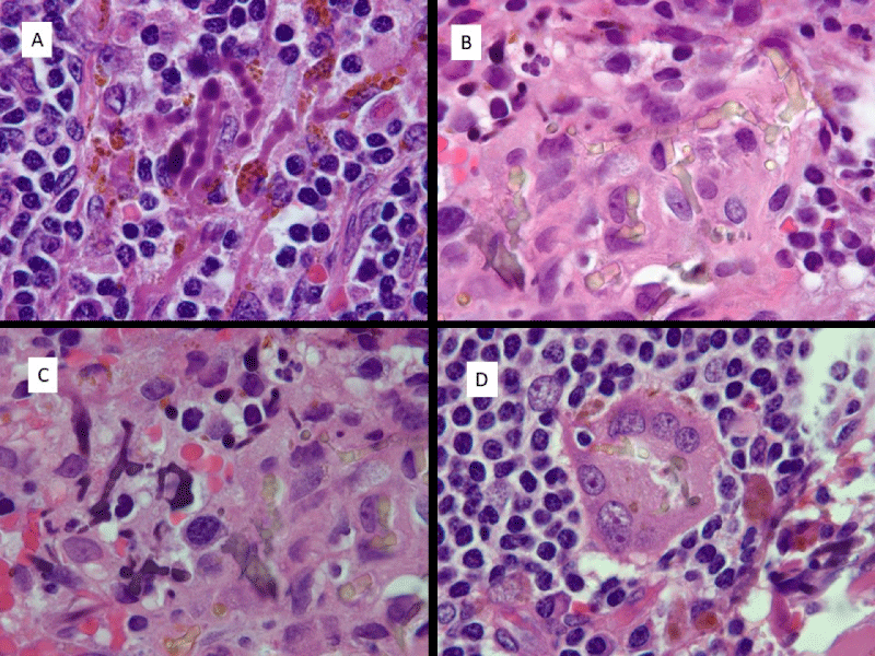
Figure 1:
Histologic appearance of pseudofungus forms. A) Basophilic, non-refractile beaded and pseudohyphal forms. B) Refractile yellowish-brown pseudohyphal forms. C) Basophilic non-refractile pseudohyphal forms on right and refractile yellowish-brown foms on left. D) Refractile pseudohyphal forms in multinucleate giant cell. (All images are hematoxylin and eosin; 1000 x magnification).
Read Full Article HTML DOI: 10.29328/journal.apcr.1001029 Cite this Article Read Full Article PDF
More Images
Similar Articles
-
MicroRNA Therapeutics in Triple Negative Breast CancerSarmistha Mitra*. MicroRNA Therapeutics in Triple Negative Breast Cancer . . 2017 doi: 10.29328/journal.hjpcr.1001003; 1: 009-017
-
Amyotropyc Lateral Sclerosis and Endogenous -Esogenous Toxicological Movens: New model to verify other Pharmacological StrategiesMauro Luisetto*,Behzad Nili-Ahmadabadi,Nilesh M Meghani,Ghulam Rasool Mashori,Ram Kumar Sahu,Kausar Rehman Khan, Ahmed Yesvi Rafa,Luca Cabianca,Gamal Abdul Hamid, Farhan Ahmad Khan. Amyotropyc Lateral Sclerosis and Endogenous -Esogenous Toxicological Movens: New model to verify other Pharmacological Strategies. . 2018 doi: 10.29328/journal.apcr.1001009; 2: 029-048
-
Receptor pharmacology and other relevant factors in lower urinary tract pathology under a functional and toxicological approach: Instrument to better manage antimicrobials therapyMauro Luisetto*,Naseer Almukhtar,Behzad Nili-Ahmadabadi,Ghulam Rasool Mashori,Kausar Rehman Khan,Ram Kumar Sahu,Farhan Ahmad Khan,Gamal Abdul Hamid,Luca Cabianca. Receptor pharmacology and other relevant factors in lower urinary tract pathology under a functional and toxicological approach: Instrument to better manage antimicrobials therapy . . 2018 doi: 10.29328/journal.apcr.1001010; 2: 049-093
-
The pathogenesis of psoriasis: insight into a complex “Mobius Loop” regulation processYuankuan Jiang,Haiyang Chen,Jiayue Liu,Tianfu Wei,Peng Ge,Jialin Qu*,Jingrong Lin. The pathogenesis of psoriasis: insight into a complex “Mobius Loop” regulation process. . 2021 doi: 10.29328/journal.apcr.1001024; 5: 020-025
-
The identification of the true nature of pseudofungus structures as polyurethane catheter fragmentsCharles M Lombard*. The identification of the true nature of pseudofungus structures as polyurethane catheter fragments. . 2022 doi: 10.29328/journal.apcr.1001029; 6: 005-008
-
Immune-mediated neuropathy related to bortezomib in a patient with multiple myelomaSusanne Koeppen*,Jörg Hense,Kay Wilhelm Nolte,Joachim Weis. Immune-mediated neuropathy related to bortezomib in a patient with multiple myeloma. . 2022 doi: 10.29328/journal.apcr.1001028; 6: 001-004
-
Post-operative agranulocytosis caused by intravenous cefazolin: A case report with a discussion of the pathogenesisCharles M Lombard*,Jiali Li,Bijayee Shrestha. Post-operative agranulocytosis caused by intravenous cefazolin: A case report with a discussion of the pathogenesis. . 2022 doi: 10.29328/journal.apcr.1001030; 6: 009-012
-
Harmonizing Artificial Intelligence Governance; A Model for Regulating a High-risk Categories and Applications in Clinical Pathology: The Evidence and some ConcernsMaxwell Omabe*. Harmonizing Artificial Intelligence Governance; A Model for Regulating a High-risk Categories and Applications in Clinical Pathology: The Evidence and some Concerns. . 2024 doi: 10.29328/journal.apcr.1001040; 8: 001-005
-
The Accuracy of pHH3 in Meningioma Grading: A Single Institution StudyMansouri Nada1, Yaiche Rahma*, Takout Khouloud, Gargouri Faten, Tlili Karima, Rachdi Mohamed Amine, Ammar Hichem, Yedeas Dahmani, Radhouane Khaled, Chkili Ridha, Msakni Issam, Laabidi Besma. The Accuracy of pHH3 in Meningioma Grading: A Single Institution Study. . 2024 doi: 10.29328/journal.apcr.1001041; 8: 006-011
Recently Viewed
-
Assessment of Albino Beech Supremacy to Pigmented Beech Proves to Be A Better Environmental Condition BioindicatorRenata Gagić-Serdar*,Miroslava Marković,Ljubinko Rakonjac,Goran Češljar,Bojan Konatar. Assessment of Albino Beech Supremacy to Pigmented Beech Proves to Be A Better Environmental Condition Bioindicator. Insights Biol Med. 2025: doi: 10.29328/journal.ibm.1001031; 9: 009-015.
-
Rare Locations of Plasma Cell Tumour: A Single-Centre ExperienceVladimir Prandjev,Donika Vezirska,Ivan Kindekov*. Rare Locations of Plasma Cell Tumour: A Single-Centre Experience. J Hematol Clin Res. 2025: doi: 10.29328/journal.jhcr.1001036; 9: 015-019
-
Age Pyramid Assessment of Commercially Important Fishes, Cirrhinus mrigala and Oreochromis niloticus, from the Tropical Yamuna River, IndiaPriyanka Mayank,Amitabh Chandra Dwivedi*. Age Pyramid Assessment of Commercially Important Fishes, Cirrhinus mrigala and Oreochromis niloticus, from the Tropical Yamuna River, India. Insights Biol Med. 2025: doi: 10.29328/journal.ibm.1001029; 9: 001-004
-
Comprehensive Acceptance Testing and Performance Evaluation of the Symbia Intevo Bold SPECT/CT System for Clinical UseSubhash Chand Kheruka*,Naema Al-Maymani,Noura Al-Makhmari,Huda Al-Saidi,Sana Al-Rashdi,Anas Al-Balushi,Anjali Jain,Khulood Al-Riyami,Rashid Al-Sukaiti. Comprehensive Acceptance Testing and Performance Evaluation of the Symbia Intevo Bold SPECT/CT System for Clinical Use. J Radiol Oncol. 2025: doi: 10.29328/journal.jro.1001076; 9: 017-030
-
Survey on the Underutilization of Forensic Expertise in India: Examining the Dominance of Law Enforcement in Evidence Collection and InvestigationsI Kanishga*,HR Bhargava. Survey on the Underutilization of Forensic Expertise in India: Examining the Dominance of Law Enforcement in Evidence Collection and Investigations. J Forensic Sci Res. 2025: doi: 10.29328/journal.jfsr.1001083; 9: 067-086
Most Viewed
-
Feasibility study of magnetic sensing for detecting single-neuron action potentialsDenis Tonini,Kai Wu,Renata Saha,Jian-Ping Wang*. Feasibility study of magnetic sensing for detecting single-neuron action potentials. Ann Biomed Sci Eng. 2022 doi: 10.29328/journal.abse.1001018; 6: 019-029
-
Evaluation of In vitro and Ex vivo Models for Studying the Effectiveness of Vaginal Drug Systems in Controlling Microbe Infections: A Systematic ReviewMohammad Hossein Karami*, Majid Abdouss*, Mandana Karami. Evaluation of In vitro and Ex vivo Models for Studying the Effectiveness of Vaginal Drug Systems in Controlling Microbe Infections: A Systematic Review. Clin J Obstet Gynecol. 2023 doi: 10.29328/journal.cjog.1001151; 6: 201-215
-
Prospective Coronavirus Liver Effects: Available KnowledgeAvishek Mandal*. Prospective Coronavirus Liver Effects: Available Knowledge. Ann Clin Gastroenterol Hepatol. 2023 doi: 10.29328/journal.acgh.1001039; 7: 001-010
-
Causal Link between Human Blood Metabolites and Asthma: An Investigation Using Mendelian RandomizationYong-Qing Zhu, Xiao-Yan Meng, Jing-Hua Yang*. Causal Link between Human Blood Metabolites and Asthma: An Investigation Using Mendelian Randomization. Arch Asthma Allergy Immunol. 2023 doi: 10.29328/journal.aaai.1001032; 7: 012-022
-
An algorithm to safely manage oral food challenge in an office-based setting for children with multiple food allergiesNathalie Cottel,Aïcha Dieme,Véronique Orcel,Yannick Chantran,Mélisande Bourgoin-Heck,Jocelyne Just. An algorithm to safely manage oral food challenge in an office-based setting for children with multiple food allergies. Arch Asthma Allergy Immunol. 2021 doi: 10.29328/journal.aaai.1001027; 5: 030-037

HSPI: We're glad you're here. Please click "create a new Query" if you are a new visitor to our website and need further information from us.
If you are already a member of our network and need to keep track of any developments regarding a question you have already submitted, click "take me to my Query."


