Figure 1
Immune-mediated neuropathy related to bortezomib in a patient with multiple myeloma
Susanne Koeppen*, Jörg Hense, Kay Wilhelm Nolte and Joachim Weis
Published: 03 January, 2022 | Volume 6 - Issue 1 | Pages: 001-004
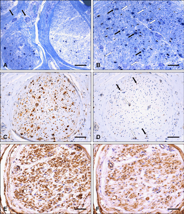
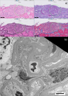
Figure 1:
Histological analysis of the sural nerve biopsy. (A) Two adjacent nerve fascicles showing severe axonal loss which is unevenly distributed throughout the fascicles and from fascicle to fascicle. Note some mononuclear inflammatory cells around small epineurial vessels (arrows) without vessel wall destruction (resin embedded tissue, semithin section, toluidine blue). (B) Semithin transverse section of a nerve fascicle with only a few myelinated nerve fibers preserved (black arrows), several acutely degenerating nerve fibers as well as myelion debris (white arrows). No hypertrophic changes (onion bulb formation), demyelinated axons or regeneration groups are seen (toluidine blue stain). (C) Numerous macrophages invading the endoneurium of a nerve fascicle, being focally clustered, e. g. around endoneurial vessels (arrows) (Paraffin section, CD68-immunostaining). (D) Cytotoxic T-lymphocytes (arrows) in the endoneurial compartment of a nerve fascicle (same as in C), sometimes closely related to vessels (white arrows) (immunohistochemical staining for the CD8 T cell surface antigen). (E, F) Mainly diffuse staining of largely equal intensity within the same nerve fascicle after incubation with antisera to immunoglobulin light chains (immunohistochemical staining for kappa, E, and lambda, F). Scale bar: (A, C, D) 90 µm, (B, E, F) 45 µm.
Read Full Article HTML DOI: 10.29328/journal.apcr.1001028 Cite this Article Read Full Article PDF
More Images
Similar Articles
-
MicroRNA Therapeutics in Triple Negative Breast CancerSarmistha Mitra*. MicroRNA Therapeutics in Triple Negative Breast Cancer . . 2017 doi: 10.29328/journal.hjpcr.1001003; 1: 009-017
-
A novel case of an infantile fibrosarcoma-like tumor with KIAA1549-BRAF translocation and an oncogenic NF2p.Q459* SNV with potential clinical significanceAnita Nagy,Consolato M Sergi,Joseph de Nanassy,Lesleigh S Abbot,Anthony Arnoldo,Cynthia Hawkins,Bo-Yee Ngan*. A novel case of an infantile fibrosarcoma-like tumor with KIAA1549-BRAF translocation and an oncogenic NF2p.Q459* SNV with potential clinical significance. . 2021 doi: 10.29328/journal.apcr.1001023; 5: 016-019
-
Immune-mediated neuropathy related to bortezomib in a patient with multiple myelomaSusanne Koeppen*,Jörg Hense,Kay Wilhelm Nolte,Joachim Weis. Immune-mediated neuropathy related to bortezomib in a patient with multiple myeloma. . 2022 doi: 10.29328/journal.apcr.1001028; 6: 001-004
Recently Viewed
-
Investigation of Stain Patterns from Diverse Blood Samples on Various SurfacesSonia Rajkumari*. Investigation of Stain Patterns from Diverse Blood Samples on Various Surfaces. J Forensic Sci Res. 2024: doi: 10.29328/journal.jfsr.1001061; 8: 028034
-
Anxiety and depression as an effect of birth order or being an only child: Results of an internet survey in Poland and GermanyJochen Hardt*,Lisa Weyer,Malgorzata Dragan,Wilfried Laubach. Anxiety and depression as an effect of birth order or being an only child: Results of an internet survey in Poland and Germany. Insights Depress Anxiety. 2017: doi: 10.29328/journal.hda.1001003; 1: 015-022
-
Pharmacovigilance is Important for Assessments of Drugs, and Withdrawal of the Drugs that have Adverse Effects More than The Benefits of Their TreatmentRezk R Ayyad,Yasser Abdel Allem Hassan,Ahmed R Ayyad*. Pharmacovigilance is Important for Assessments of Drugs, and Withdrawal of the Drugs that have Adverse Effects More than The Benefits of Their Treatment. Arch Pharm Pharma Sci. 2025: doi: 10.29328/journal.apps.1001069; 9: 042-045
-
The Impact of Artificial Intelligence on the Daily Responsibilities of Family Doctors: A Comprehensive Review of Current KnowledgeAdawi Mohammad*,Awni Yousef. The Impact of Artificial Intelligence on the Daily Responsibilities of Family Doctors: A Comprehensive Review of Current Knowledge. J Community Med Health Solut. 2025: doi: 10.29328/journal.jcmhs.1001061; 6: 067-076
-
Forensic Psychology and Criminal ProfilingEze SM*,Alabi KJ,Yusuf AO,Hamzat FO,A Abdulrauf,Atoyebi AT,Lawal IA,OA Ibrahim,AY Imam-Fulani,Dare BJ. Forensic Psychology and Criminal Profiling. J Forensic Sci Res. 2025: doi: 10.29328/journal.jfsr.1001085; 9: 092-096
Most Viewed
-
Feasibility study of magnetic sensing for detecting single-neuron action potentialsDenis Tonini,Kai Wu,Renata Saha,Jian-Ping Wang*. Feasibility study of magnetic sensing for detecting single-neuron action potentials. Ann Biomed Sci Eng. 2022 doi: 10.29328/journal.abse.1001018; 6: 019-029
-
Evaluation of In vitro and Ex vivo Models for Studying the Effectiveness of Vaginal Drug Systems in Controlling Microbe Infections: A Systematic ReviewMohammad Hossein Karami*, Majid Abdouss*, Mandana Karami. Evaluation of In vitro and Ex vivo Models for Studying the Effectiveness of Vaginal Drug Systems in Controlling Microbe Infections: A Systematic Review. Clin J Obstet Gynecol. 2023 doi: 10.29328/journal.cjog.1001151; 6: 201-215
-
Prospective Coronavirus Liver Effects: Available KnowledgeAvishek Mandal*. Prospective Coronavirus Liver Effects: Available Knowledge. Ann Clin Gastroenterol Hepatol. 2023 doi: 10.29328/journal.acgh.1001039; 7: 001-010
-
Causal Link between Human Blood Metabolites and Asthma: An Investigation Using Mendelian RandomizationYong-Qing Zhu, Xiao-Yan Meng, Jing-Hua Yang*. Causal Link between Human Blood Metabolites and Asthma: An Investigation Using Mendelian Randomization. Arch Asthma Allergy Immunol. 2023 doi: 10.29328/journal.aaai.1001032; 7: 012-022
-
An algorithm to safely manage oral food challenge in an office-based setting for children with multiple food allergiesNathalie Cottel,Aïcha Dieme,Véronique Orcel,Yannick Chantran,Mélisande Bourgoin-Heck,Jocelyne Just. An algorithm to safely manage oral food challenge in an office-based setting for children with multiple food allergies. Arch Asthma Allergy Immunol. 2021 doi: 10.29328/journal.aaai.1001027; 5: 030-037

HSPI: We're glad you're here. Please click "create a new Query" if you are a new visitor to our website and need further information from us.
If you are already a member of our network and need to keep track of any developments regarding a question you have already submitted, click "take me to my Query."


