Figure 2
Uterine precursor lesions in patients with incidental nodal lymphangioleiomyomatosis: A report of 4 cases
Charles M Lombard*
Published: 14 December, 2020 | Volume 4 - Issue 1 | Pages: 001-004.

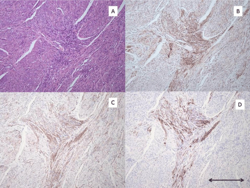
Figure 2:
Pre-LAM uterine lesion pattern #2 A): H/E stain of area with pre-LAM lesion composed of spindle cells without distinct morphologic variance from surrounding uterine smooth muscle cells. B): Cathepsin K IHC stain with strong positive staining of the small stellate proliferation of pre-LAM cells. C): Beta-catenin IHC stain with moderate positive staining of the small stellate proliferation of pre-LAM cell. D): HMB-45 IHC stain with strong positive staining of the small stellate proliferaton of pre-LAM cells. All photomicrographs taken at 100x magnification. Scale bar in lower right corner measures 0.2 mm.
Read Full Article HTML DOI: 10.29328/journal.apcr.1001016 Cite this Article Read Full Article PDF
More Images
Similar Articles
-
MicroRNA Therapeutics in Triple Negative Breast CancerSarmistha Mitra*. MicroRNA Therapeutics in Triple Negative Breast Cancer . . 2017 doi: 10.29328/journal.hjpcr.1001003; 1: 009-017
-
Amyotropyc Lateral Sclerosis and Endogenous -Esogenous Toxicological Movens: New model to verify other Pharmacological StrategiesMauro Luisetto*,Behzad Nili-Ahmadabadi,Nilesh M Meghani,Ghulam Rasool Mashori,Ram Kumar Sahu,Kausar Rehman Khan, Ahmed Yesvi Rafa,Luca Cabianca,Gamal Abdul Hamid, Farhan Ahmad Khan. Amyotropyc Lateral Sclerosis and Endogenous -Esogenous Toxicological Movens: New model to verify other Pharmacological Strategies. . 2018 doi: 10.29328/journal.apcr.1001009; 2: 029-048
-
Receptor pharmacology and other relevant factors in lower urinary tract pathology under a functional and toxicological approach: Instrument to better manage antimicrobials therapyMauro Luisetto*,Naseer Almukhtar,Behzad Nili-Ahmadabadi,Ghulam Rasool Mashori,Kausar Rehman Khan,Ram Kumar Sahu,Farhan Ahmad Khan,Gamal Abdul Hamid,Luca Cabianca. Receptor pharmacology and other relevant factors in lower urinary tract pathology under a functional and toxicological approach: Instrument to better manage antimicrobials therapy . . 2018 doi: 10.29328/journal.apcr.1001010; 2: 049-093
-
Uterine precursor lesions in patients with incidental nodal lymphangioleiomyomatosis: A report of 4 casesCharles M Lombard*. Uterine precursor lesions in patients with incidental nodal lymphangioleiomyomatosis: A report of 4 cases . . 2020 doi: 10.29328/journal.apcr.1001016; 4: 001-004.
-
The pathogenesis of psoriasis: insight into a complex “Mobius Loop” regulation processYuankuan Jiang,Haiyang Chen,Jiayue Liu,Tianfu Wei,Peng Ge,Jialin Qu*,Jingrong Lin. The pathogenesis of psoriasis: insight into a complex “Mobius Loop” regulation process. . 2021 doi: 10.29328/journal.apcr.1001024; 5: 020-025
-
Immune-mediated neuropathy related to bortezomib in a patient with multiple myelomaSusanne Koeppen*,Jörg Hense,Kay Wilhelm Nolte,Joachim Weis. Immune-mediated neuropathy related to bortezomib in a patient with multiple myeloma. . 2022 doi: 10.29328/journal.apcr.1001028; 6: 001-004
-
Post-operative agranulocytosis caused by intravenous cefazolin: A case report with a discussion of the pathogenesisCharles M Lombard*,Jiali Li,Bijayee Shrestha. Post-operative agranulocytosis caused by intravenous cefazolin: A case report with a discussion of the pathogenesis. . 2022 doi: 10.29328/journal.apcr.1001030; 6: 009-012
-
Harmonizing Artificial Intelligence Governance; A Model for Regulating a High-risk Categories and Applications in Clinical Pathology: The Evidence and some ConcernsMaxwell Omabe*. Harmonizing Artificial Intelligence Governance; A Model for Regulating a High-risk Categories and Applications in Clinical Pathology: The Evidence and some Concerns. . 2024 doi: 10.29328/journal.apcr.1001040; 8: 001-005
-
The Accuracy of pHH3 in Meningioma Grading: A Single Institution StudyMansouri Nada1, Yaiche Rahma*, Takout Khouloud, Gargouri Faten, Tlili Karima, Rachdi Mohamed Amine, Ammar Hichem, Yedeas Dahmani, Radhouane Khaled, Chkili Ridha, Msakni Issam, Laabidi Besma. The Accuracy of pHH3 in Meningioma Grading: A Single Institution Study. . 2024 doi: 10.29328/journal.apcr.1001041; 8: 006-011
Recently Viewed
-
Acute Gas Toxicity at Work: A Tale of Two Cases with Review of LiteratureRishabh Kumar Singh,Jitender Pratap Singh,Manjari Kishore*,HM Garg. Acute Gas Toxicity at Work: A Tale of Two Cases with Review of Literature. J Forensic Sci Res. 2025: doi: 10.29328/journal.jfsr.1001091; 9: 125-128
-
Minds after Death: The Expanding Role of Psychological Autopsy in Investigations: A ReviewIshan Jain*,Oindrila Mahapatra,Yogesh Kumar. Minds after Death: The Expanding Role of Psychological Autopsy in Investigations: A Review. J Forensic Sci Res. 2025: doi: 10.29328/journal.jfsr.1001096; 9: 155-0
-
Role of Perianesthesia Nurses in Enhanced Recovery After Surgery (ERAS) Protocols: A Narrative Review and Comparative Outcomes AnalysisOghogho Linda Akarogbe*,Geneva Igwama,Olachi Lovina Emenyonu,Idowu M Ariyibi. Role of Perianesthesia Nurses in Enhanced Recovery After Surgery (ERAS) Protocols: A Narrative Review and Comparative Outcomes Analysis. Int J Clin Anesth Res. 2025: doi: 10.29328/journal.ijcar.1001034; 9: 037-039
-
Brain washing systems and other circulating factors in some neurological condition like Parkinson (Pd) and vascular and diabetic dementia: How dynamics- saturation of clearance can act on toxic molecule?Mauro Luisetto*,Farhan Ahmad Khan,Akram Muhamad,Ghulam Rasool Mashori,Behzad Nili Ahmadabadi,Oleg Yurevich Latiyshev. Brain washing systems and other circulating factors in some neurological condition like Parkinson (Pd) and vascular and diabetic dementia: How dynamics- saturation of clearance can act on toxic molecule?. J Neurosci Neurol Disord. 2020: doi: 10.29328/journal.jnnd.1001028; 4: 001-013
-
Do genes matter in sleep?-A comprehensive updateRajib Dutta*. Do genes matter in sleep?-A comprehensive update. J Neurosci Neurol Disord. 2020: doi: 10.29328/journal.jnnd.1001029; 4: 014-023
Most Viewed
-
Feasibility study of magnetic sensing for detecting single-neuron action potentialsDenis Tonini,Kai Wu,Renata Saha,Jian-Ping Wang*. Feasibility study of magnetic sensing for detecting single-neuron action potentials. Ann Biomed Sci Eng. 2022 doi: 10.29328/journal.abse.1001018; 6: 019-029
-
Evaluation of In vitro and Ex vivo Models for Studying the Effectiveness of Vaginal Drug Systems in Controlling Microbe Infections: A Systematic ReviewMohammad Hossein Karami*, Majid Abdouss*, Mandana Karami. Evaluation of In vitro and Ex vivo Models for Studying the Effectiveness of Vaginal Drug Systems in Controlling Microbe Infections: A Systematic Review. Clin J Obstet Gynecol. 2023 doi: 10.29328/journal.cjog.1001151; 6: 201-215
-
Prospective Coronavirus Liver Effects: Available KnowledgeAvishek Mandal*. Prospective Coronavirus Liver Effects: Available Knowledge. Ann Clin Gastroenterol Hepatol. 2023 doi: 10.29328/journal.acgh.1001039; 7: 001-010
-
Causal Link between Human Blood Metabolites and Asthma: An Investigation Using Mendelian RandomizationYong-Qing Zhu, Xiao-Yan Meng, Jing-Hua Yang*. Causal Link between Human Blood Metabolites and Asthma: An Investigation Using Mendelian Randomization. Arch Asthma Allergy Immunol. 2023 doi: 10.29328/journal.aaai.1001032; 7: 012-022
-
An algorithm to safely manage oral food challenge in an office-based setting for children with multiple food allergiesNathalie Cottel,Aïcha Dieme,Véronique Orcel,Yannick Chantran,Mélisande Bourgoin-Heck,Jocelyne Just. An algorithm to safely manage oral food challenge in an office-based setting for children with multiple food allergies. Arch Asthma Allergy Immunol. 2021 doi: 10.29328/journal.aaai.1001027; 5: 030-037

HSPI: We're glad you're here. Please click "create a new Query" if you are a new visitor to our website and need further information from us.
If you are already a member of our network and need to keep track of any developments regarding a question you have already submitted, click "take me to my Query."


